Radiographic Testing (RT) for Pressure Vessels: A Comprehensive Guide
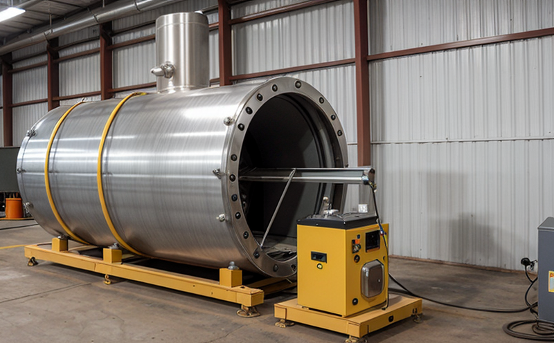
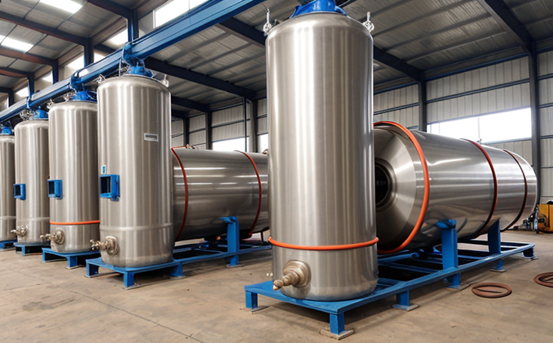
Pressure vessels are critical components in various industries, including chemical processing, petroleum refining, and power generation. These vessels contain fluids or gases under pressure, and their integrity is essential to prevent catastrophic failures that can lead to injuries, environmental damage, and costly repairs. Radiographic testing (RT) is a non-destructive testing (NDT) method used to inspect the welds and materials of pressure vessels for defects, such as cracks, voids, or inclusions.
Radiographic testing involves the use of ionizing radiation to produce an image of the internal structure of the vessel. The process typically consists of exposing the vessels welds to X-rays or gamma rays, which penetrate the material and create a radiograph. A radiograph is essentially an X-ray image that reveals the internal defects within the material. The resulting image can be analyzed by trained personnel to detect any anomalies.
There are several types of RT methods used for pressure vessels, including:
Film radiography (FILM): This traditional method uses film as the detector medium to capture the radiograph.
Digital radiography (DR): Also known as computed radiography (CR), this method uses a digital detector to produce a radiographic image.
Computed tomography (CT) scanning: Although not typically used for pressure vessel inspection, CT scanning can be employed in specific cases where detailed, three-dimensional imaging is required.
Radiographic Testing Equipment and Procedures
Radiographic testing equipment consists of several components:
X-ray or gamma radiation source
Detector medium (film or digital)
Intensifying screens (for film radiography)
Processing chemicals (for film radiography)
The RT process typically follows these steps:
1. Prepare the vessel for inspection: Remove any non-essential equipment, cover exposed areas with lead sheeting, and ensure access to all welds.
2. Position the detector medium: Place the film or digital detector near the area of interest, taking care not to obscure the radiographic image.
3. Expose the vessel: Direct the X-ray or gamma radiation source at the welds, using a collimator to focus the beam and reduce scatter.
4. Process the radiograph (for FILM): Develop the film using intensifying screens and processing chemicals.
Digital Radiography: An Overview
Digital radiography offers several advantages over traditional film radiography:
Higher sensitivity
Faster image acquisition times
Reduced exposure levels
Improved image quality
Some key features of digital radiography include:
Detector types: Digital detectors can be categorized into two main groups:
Flat panel detectors (FPD): These use a solid-state technology to capture X-ray photons.
Computed radiography (CR) phosphor plate: This type uses a photostimulable phosphor plate that captures the radiographic image.
Image acquisition: Digital radiography can be performed using various methods:
Direct exposure
Indirect exposure
Computed tomography (CT)
Computed Tomography Scanning for Pressure Vessels
Although not a standard RT method, CT scanning has been used in specific applications where high-resolution images are necessary:
Advantages: CT scanning offers excellent image quality and can detect small defects.
Disadvantages: The process is complex, requires specialized equipment, and involves higher costs.
Some common uses of CT scanning for pressure vessels include:
Void detection
Inclusion analysis
Surface crack inspection
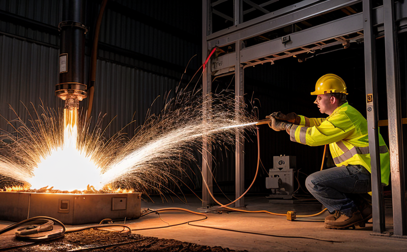
Quality Control in Radiographic Testing
The accuracy and reliability of RT results rely heavily on proper quality control measures. Some essential steps to ensure quality control in RT include:
Proper calibration of equipment
Regular maintenance of testing devices
Training and certification for personnel involved in the process
Radiographic Testing Limitations and Considerations
While radiographic testing is a valuable tool for pressure vessel inspection, it has some limitations:
Detection sensitivity: Radiography may not detect very small defects or those located near the surface.
Image interpretation: Trained analysts are required to interpret the results accurately.
To mitigate these limitations, RT should be used in conjunction with other NDT methods, such as ultrasonic testing (UT) and magnetic particle inspection (MPI).
QA Section
1. What types of pressure vessels can benefit from radiographic testing?
Answer: All types of pressure vessels, including spherical, cylindrical, and ellipsoidal shapes.
2. How often should radiographic testing be performed on pressure vessels?
Answer: The frequency of RT depends on various factors, such as vessel operating conditions, material type, and inspection history. Typically, RT is performed every 5-10 years or after a certain number of cycles.
3. Can radiographic testing detect defects in composite materials?
Answer: Yes, but with limitations. Radiography can detect voids and delamination within the material.
4. What are some common sources of error in radiographic testing?
Answer: Sources include inadequate equipment calibration, incorrect exposure settings, and insufficient training for personnel involved in the process.
5. Can I perform radiographic testing on a vessel that has been repaired or modified since its initial inspection?
Answer: Yes, but its essential to follow proper procedures to ensure that the repair or modification does not affect the accuracy of the test results.
6. How do I ensure accurate interpretation of radiographic images?
Answer: Trained analysts should interpret the results using a combination of visual examination and comparison with reference standards.
7. Can digital radiography be used for inspection of complex vessel geometries?
Answer: Yes, but it may require specialized equipment or techniques to capture high-quality images.
8. What are some common applications of CT scanning in pressure vessel inspection?
Answer: Void detection, inclusion analysis, surface crack inspection, and 3D imaging for detailed analysis.
9. Can radiographic testing detect defects that occur during the manufacturing process?
Answer: Yes, but only if they are present at a sufficient size to be detected by RT.
10. How do I ensure compliance with industry standards for radiographic testing of pressure vessels?
Answer: Familiarize yourself with relevant codes and standards, such as ASME Boiler and Pressure Vessel Code (BPVC) Section V, and follow proper procedures for equipment calibration, testing, and image interpretation.